 Overview
Vision is an important way for interacting with our world. Take it from someone who has low vision that not being able to see well is very inconvenient.
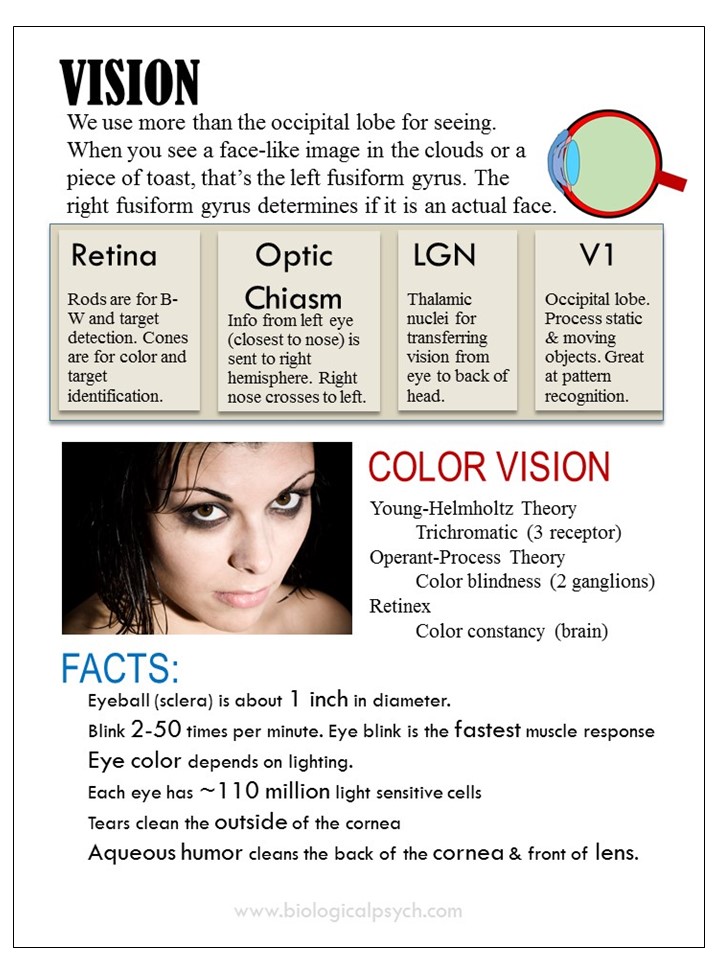
The eye is a transducer of light into neural impulses. Light hits the cornea (the clear dome cover on the front of the eye) and is focused on the lens. The cornea provides two-thirds of the eye’s focus. It’s just not an adjustable focus.
Light shines through the pupil of the iris, hits the lens (of its one-third adjustment) and travels to the retina to be processed. From then on, it is all about neuron connections and processing.
Pay particular attention to visual coding, summation and color vision.
Learning Objectives
By the end of this module, you should be able to:
- Describe how cells in the occipital lobe process shape and movement
- Explain the difference between the scotopic and photic systems
- Compare and contrast the dorsal and temporal pathways
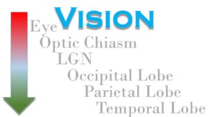
- Describe the path from the eye to the occipital lobe
- Compare and contrast rods and cones
Readings
- Kalat C5
- Eye & Retina
- Occipital Lobe
Slides
Videos
Discussion
If you went blind, what would you miss the most and what would stay pretty much the same?
Quiz (not the same as on Canvas)
Written Assignment
- Describe the three (3) types of cones and what they do.
- Compare the temporal and dorsal steams. If one is damaged, what will you be able to do and not do?
- What did you find difficult or confusing in this chapter? If nothing was difficult or confusing, what did you find most interesting?
Study Guide
Key Terms
- astigmatism
- binocular
- bipolar cells
- blind spot
- blindsight
- color constancy
- color vision deficiency
- complex cells
- cones
- dorsal stream
- end-stopped (hypercomplex) cells
- feature detectors
- fovea
- fusiform gyrus
- ganglion cells
- horizontal cells
- interior temporal cortex
- koniocellular neurons
- lateral inhibition
- latergal geniculate nucleus (LGN)
- law of specific nerve energies
- magnocellular neurons
- midget ganglion cells
- motion blindness
- MST
- MT (area V5)
- negative color afterimage
- opponent-process theory
- optic nerve
- parvocellular neurons
- photopigment
- primary visual cortex (area V1)
- prosopagnosia
- pupil
- receptive field
- retina
- retinal disparity
- retinex theory
- rods
- saccade
- secondary visual cortex
- sensitive period
- simple cell
- stabismus
- trichromatic theory (Young-Helmholtz theory)
- ventral stream
- visual agnosia
- visual field
Links
- Neuroscience for Kids: The Blind Spot (Links to an external site.)
- Neuroscience for Kids: The Retina (Links to an external site.)
- DrugAbuse.gov’s post on Drugs, Brains & Behaviors (Links to an external site.)
- Neuroscience for Kids: Alcohol (Links to an external site.)
Summary
We use more than the occipital lobe for seeing. When you can see a face-like image in the clouds or a piece of toast, that’s the left fusiform gyrus. The right fusiform gyrus determines if it is an actual face.